The group collaborates closely with the Wolfson Brain Imaging Centre at Addenbrookes Hospital to develop improved methods for Magnetic Resonance Imaging (MRI) and Positron Emission Tomography (PET). The joint projects include Algorithm development to improve clinical utility, and novel hardware development.
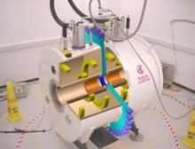
Combined PET-MRI scanner
Positron emission tomography is a nuclear medicine medical imaging technique which produces a three-dimensional image or map of functional processes in the body. The system detects pairs of gamma rays emitted indirectly by a positron-emitting radioisotope, which is introduced into the body on a metabolically active molecule. Positron emission tomography is primarily used in oncology and to detect diseases of the brain and heart.
Magnetic resonance imaging is primarily used in medical imaging to visualise the structure and function of the body. It provides detailed images of the body in three dimensions. The scanner creates a powerful magnetic field which aligns the magnetization of hydrogen atoms in the body. Radio waves are used to alter the alignment of this magnetization. This causes the hydrogen atoms to emit a weak radio signal which is amplified by the scanner. This signal can be manipulated by additional magnetic fields to build up enough information to reconstruct an image of the body.
Combined use of these two imaging techniques allows researchers to observe simultaneously the functional and structural modes of organs, and also serves to confirm specific tissue functions using the two complementary observations. The PET-MRI development is based in the Cavendish where there is a 1 Tesla superconducting MRI magnet with a room temperature bore of 35 cm. The instrument is now installed at the new Pre-clinical Imaging Centre in the Clinical school at Addenbrookes.