Most of what we know in Physics has been derived from experience with the inanimate world. It is a challenge to transfer these concepts to living objects such as cells, tissues, and entire organisms, where it is not certain if they are appropriate or even relevant. The group investigates the mechanical and optical properties of living cells and tissues using novel photonic tools to test their relevance and importance for biological function. The ultimate goal is the transfer of findings to medical application in the fields of improved diagnosis of diseases and novel approaches in regenerative medicine with an impact on clinical practice.
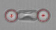
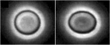 BSS combines a unique set of techniques to study the mechanical properties of cells. It includes active (optical tweezers) and passive (particle tracking) microrheology, optical stretcher, micropipette aspiration, magnetic tweezers, and atomic force microscopy (AFM). We are interested in the relation between structure (membrane, cytoskeleton, nucleus) and mechanical properties, and their relation to physiological and pathological processes. For example, changes in the cytoskeleton are diagnostic for cancer and differentiation of stem cells can be sensitively monitored by mechanical signature.
Scientists have been puzzled for ages why the vertebrate retina is inverted - the light sensitive cells are on the wrong side. Light has to traverse several hundred wavelengths of scattering tissue before it is detected with potentially detrimental effects on image quality. Recently we found that elongated glial cells in the vertebrate retina act as optical fibres channeling light through otherwise scattering layers. Their parallel array in the retina is reminiscent of fibre optic plates used for low distortion image transfer, thus suggesting a similar function in vertebrate vision.
 The paradigm that neurons in the central nervous system cannot regenerate is gone. While most research to date is biochemical, we are developing tools to investigate axonal growth and to identify mechanical cues underlying axonal behaviour. Specifically, we investigate the mechanical properties of glial scars, inevitably occurring after neurological trauma, with an AFM in order to find out whether those pose mechanical barriers to nerve regeneration. Investigation into the importance of mechanical cues for normal differentiation and axonal path finding during development and for improved acceptance of foreign implants is also ongoing.
Understanding the chemical and physical factors which control cell-substrate interaction is crucial for both fundamental and applied studies, including the development of prosthetic devices and tissue scaffolds. We are studying the effect of modifying substrates with chemical and topographical patterns to explore cell adhesion, spreading and proliferation.