Winners announced for the Cavendish Photography Competition 2026
15 October 2026
The 2026 Cavendish Photography Competition showcased a captivating collection of images celebrating this year’s theme, ‘Big and Small: exploring the breadth of Physics at the Cavendish’.
We received an overwhelming response to this year’s call, where entries offered fascinating perspectives on the extraordinary range of scales across which physics shapes our understanding of the world. Reflecting the breadth and diversity of physics research at the Cavendish, the submissions explored the beauty, complexity and connections between the microscopic and macroscopic world.
The competition invited staff, researchers and students to interpret what ‘Big and Small’ means to them through photography and visual storytelling. The resulting entries captured not only the scientific significance of their subjects but also the creativity and artistry that bring physics to life.
The winning photographs reflect the Department’s vibrant and interdisciplinary research culture, spanning areas such as Biological Physics, High Energy Physics, Energy Materials, Physics of Soft Matter and NanoSystems, and Astrophysics.
All entries were judged anonymously on the basis of thematic relevance, uniqueness and visual appeal. The judging panel considered how effectively each submission interpreted this year’s theme while offering an original and engaging perspective on physics research.
The panel of judges for this year’s Photography Competition included Prof Mete Atature, Head of the Cavendish Laboratory; Prof Siân Dutton, Deputy Head of Department; David Cain, University of Cambridge Public Engagement and Festivals Manager; Dr Sandro Tacchella, Associate Professor of Extragalactic Astrophysics; Emily Hogg, Research Laboratory Manager (Ray Dolby Centre); Minerva Maheshwari, PhD student in High Energy Physics.
We are delighted to announce the 2026 winners:
Head of Department Prize
Awarded to postdoctoral researcher Abhirup Mookherjee from Fusco Lab for both his submissions.
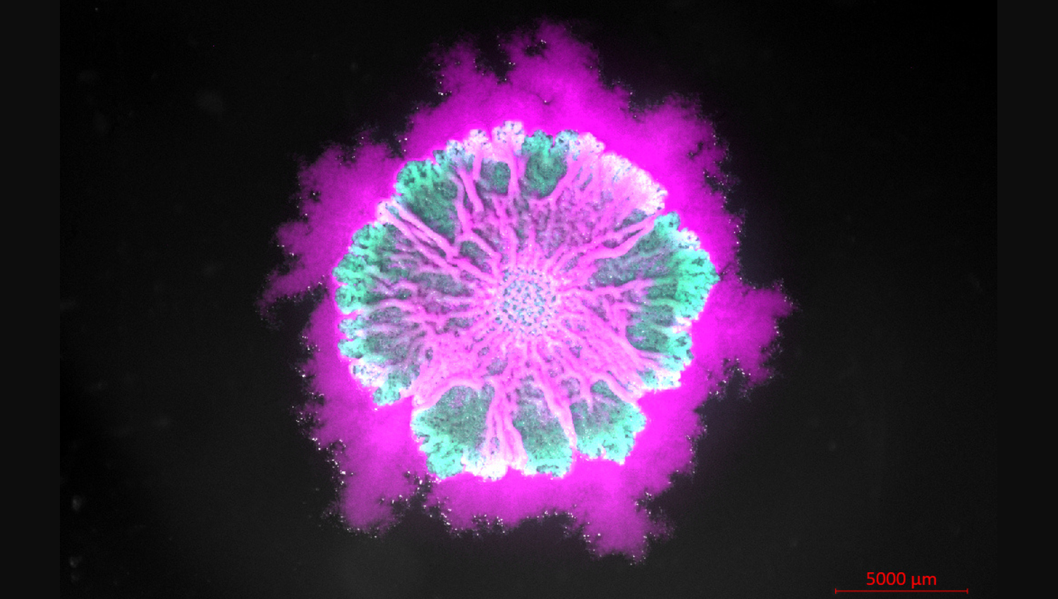
Bacterial Art in Bloom: The swimmers, the builders, and the living tapestry
What happens when two flawed microbes are forced to live together? In this vibrant biofilm image, two defective strains of Bacillus subtilis unite to build a thriving community: glowing magenta “swimmers” (which can move but lack sticky glue) and glowing green “builders” (which produce the protective matrix glue but cannot move). Overlaid with standard light microscopy, the image captures their microscopic spatial dance. Through localized growth and spatial crowding, the two populations drive the colony to fold and buckle into stunning 3D patterns across the surface.
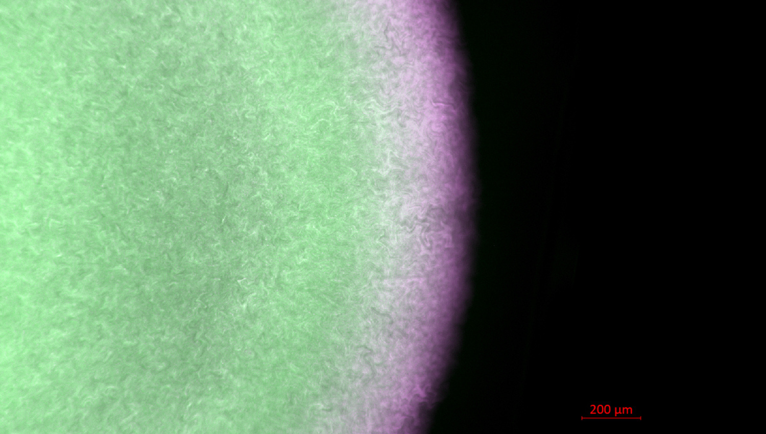
A Division of Microscopic Labor: How single bacteria team up to build a community
Even simple microscopic bacteria work as a team to build complex living communities. In this vibrant snapshot of a day-old Escherichia coli colony, different jobs are assigned to distinct zones: glowing green highlights the “builders” weaving a protective glue to anchor the colony, while magenta marks the “swimmers” forming an outer ring prepared for movement. A closer look reveals individual cells chaining, dividing, and jostling against one another. As single microbes crowd, push, and cooperate at the microscopic level, they create striking, ring-like patterns that lay the template for multicellular biofilm development.
Acknowledgements: Physics of Medicine and Fusco Lab
First Prize
Awarded to PhD student Zofia Dziekan from NanoPhotonics.
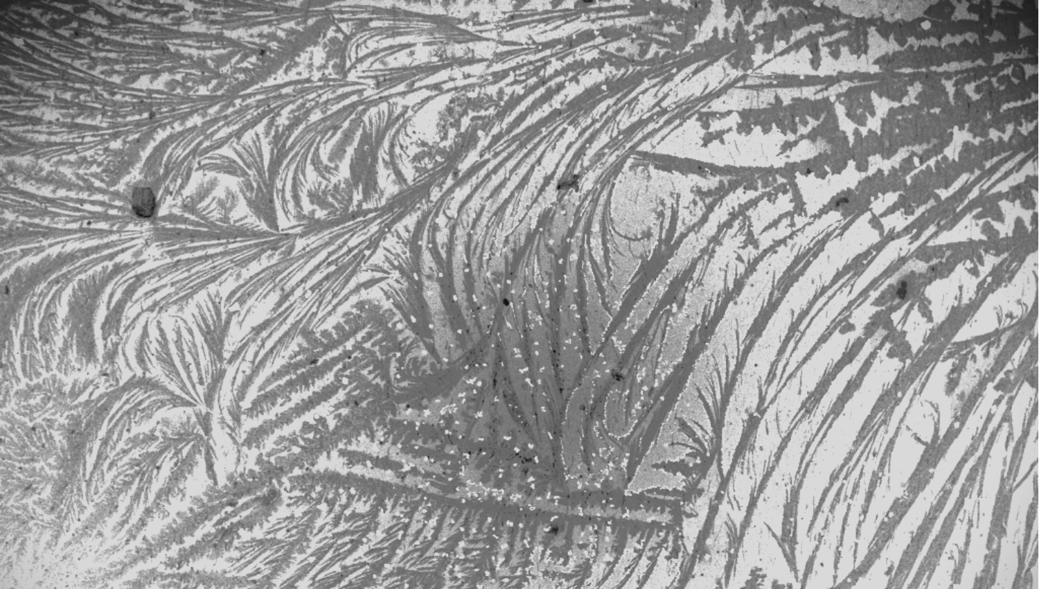
Winter Wonderland
The entry speaks to this year’s theme, Big and Small, through the story of how matter organises itself across scales. Each piece begins with nanoparticles only hundreds of nanometres in size, which then assemble into ordered sheets, lattices, or films large enough to see with the naked eye. Together, they show how structures born in the nanoscale world can grow into striking macroscopic patterns.
This scanning electron microscope image, Winter Wonderland, shows a monolayer of gold nanoparticles assembled using the linker molecule cucurbit[5]uril. During drying, the linker crystallised on the surface, creating intricate, fractal‑like patterns that give the scene its “winter wonderland” appearance. After imaging, only brightness and contrast were adjusted to make the structures more visible.
Acknowledgement: Dr Jin Qin for preparing the sample.
Second Prize
Awarded to undergraduate student Izzy Sargent.
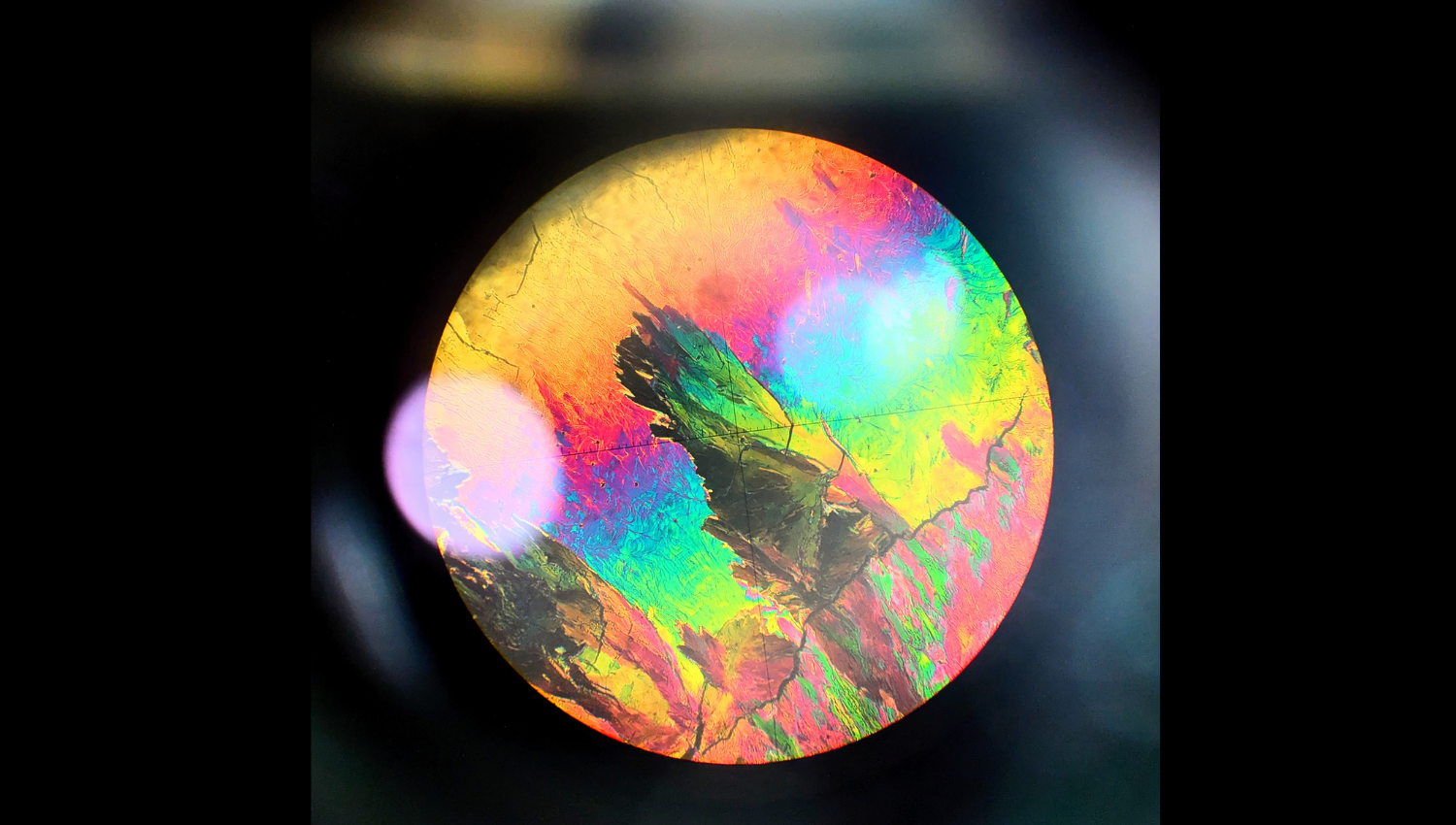
Small Made Big
Observations of liquid crystals between crossed polarisers elegantly demonstrate how nanoscale differences in molecular orientation and interactions produce macroscopic functional and visual differences. Since they inhabit the phase space between an isotropic liquid and a highly ordered solid, behaviour at the macroscopic level is a direct, visible consequence of small-scale changes.
When viewed through crossed polarisers, the discrete nature of the molecules and their alignment can be abstracted away, and we can view the liquid crystal system as a continuous medium with varying birefringent properties. Manipulation of the polarization of light provides the foundation for various technologies, most famously liquid crystal displays (LCDs). Here, nanoscale molecular orientation (small) is translated to visual information (big).
This photo of the crystalline phase of 4-n-heptyloxybenzoic acid was taken through the eyepiece of a polarised optical microscope while characterising the phase diagram of the nOBA (4-n-alkoxybenzoic acid) homologues. In this series, the lower homologues (n = 1, 2) are non-mesogenic, while intermediate chains (n = 3 to 6) show nematic liquid crystal behaviour, and longer chains (n ≥ 7) additionally exhibit smectic phases.
The image doubly captures this year’s theme “Big and Small”. We observe the “small” microscale sample section that has been magnified, revealing the “big” visual display. Equally, the “big” patterns that we see directly result from the “small” nanoscale molecular interactions and alignment.
Third Prize
Awarded to PhD student Ella Wood from High Energy Physics.
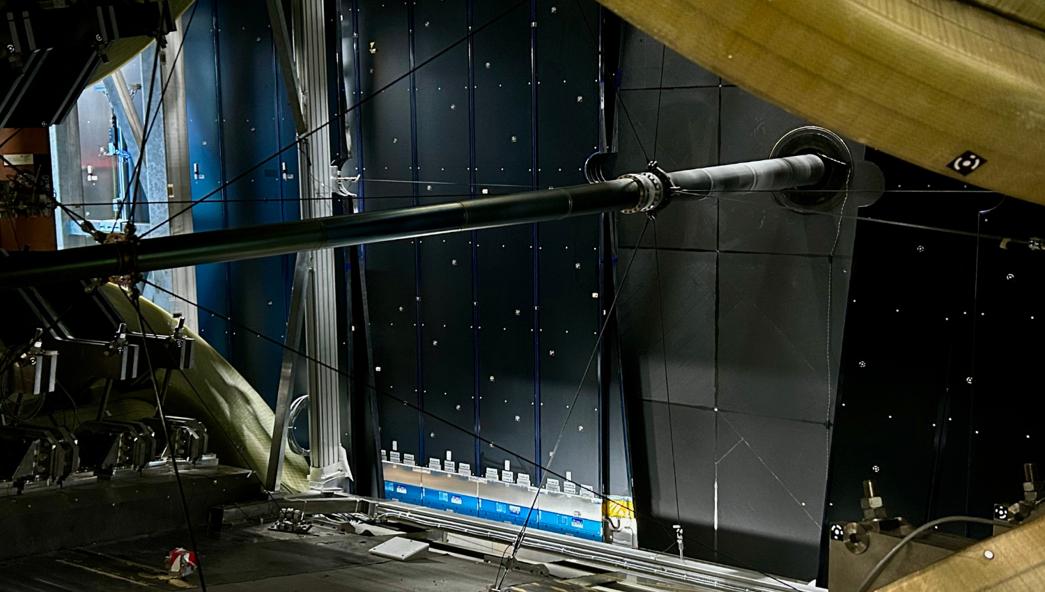
Journey to the Big Bang
Peering through the heart of the LHCb detector’s immense dipole magnet, we glimpse the SciFi subdetector used to track the paths of charged particles produced in collisions of either protons or lead ions at CERN’s Large Hadron Collider. Here it is transformed into a starry sky by the countless reflective stickers used to precisely align the subdetector, yielding a shimmering reminder of this gigantic machine’s astonishing sensitivity.
As lead ions fly through the suspended beryllium beampipe towards their collision point, they embark on a journey to recreate the conditions found just a fraction of a second after the Big Bang, enabling us to better understand the type of matter (quark-gluon plasma) that filled the early Universe. By probing this fleeting moment with high-precision measurements of the tiny particles produced in LHC collisions, we trace the evolution of matter in the wake of the Big Bang.

The text in this work is licensed under a Creative Commons Attribution-NonCommercial-ShareAlike 4.0 International License. Images, including our videos, are Copyright ©University of Cambridge and licensors/contributors as identified. All rights reserved. We make our image and video content available in a number of ways – on our main website under its Terms and conditions, and on a range of channels including social media that permit your use and sharing of our content under their respective Terms.